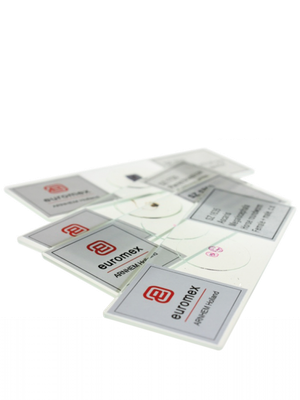
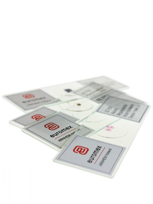
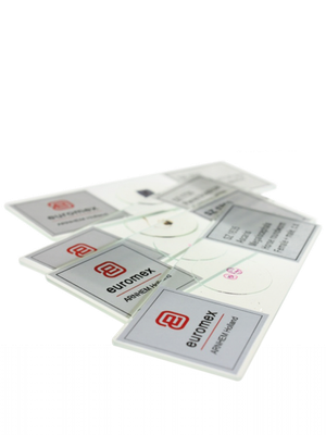
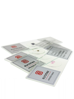
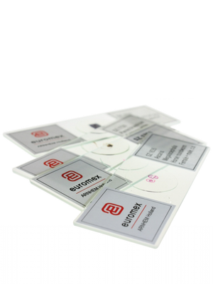
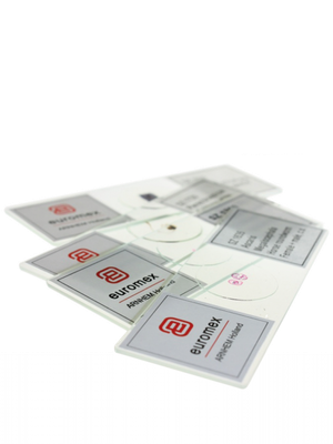
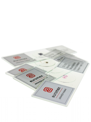
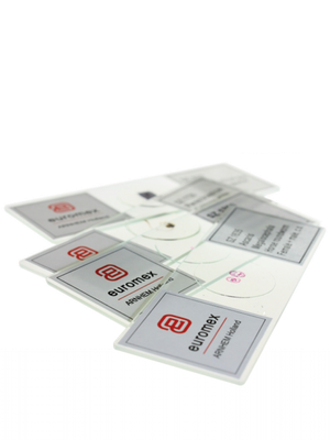
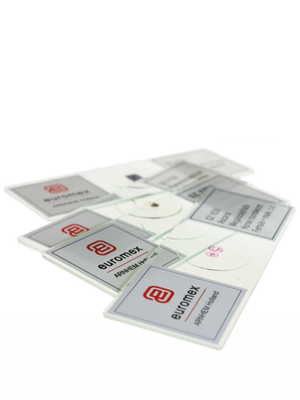
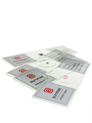
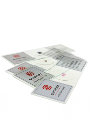
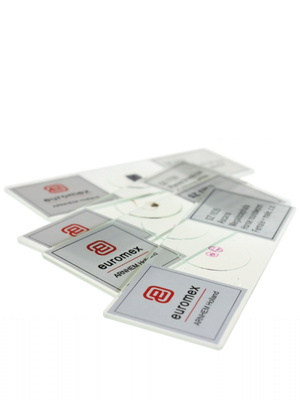
Micropreparations set 1 (25 pieces)
[tab name='Description']
Euromex offers both teachers and microscopy amateurs a wide range of quality preparation sets. These sets can be used directly in biology lessons or as an introduction to zoology and botany.
Series with 25 micropreparations for human & mammalian histology, zoology and botany. Each set contains a variety of micro preparations (see details in next tab) All micro preparations are supplied in a plastic preparation box for 25 preparations
This set contains:
Histology
SH.1011 Bone tissue human, section
SH.1045 Skeletal muscle dog, ls and cs
SH.1072 Skin with hair follicle, human
SH.1078 Skin, transverse section with different layers, dog
SH .1150 Human blood smear, Giemsa stain
Zoology
SZ.1510 Amoeba, Amoeba proteus, wm
SZ.1580 Freshwater polyp, Hydra, with budding, wm
SZ .1640 Horse roundworm, Ascaris megalocephala, mitotic eggs
SZ.1655 Waterflo, Daphnia sp., wm
SZ.1715 Housefly, Musca domestica, mouthparts
SZ.1717 Housefly, Musca domestica, leg with hooks
SZ.1719 Housefly, Musca domestica, wing, wm
Pantkunde
SB.2006 Onion, Allium cepa, scale epidermis wm
SB.2009 Onion, Allium cepa, mitosis root tip ls
SB.2055 Maize, Zea mays, stem, cs
SB.2060 Wheat, Triticumaestivum, stem, cs
SB.2070 Sunflower, Helianthus, young stem with main bundles, cs
SB. 2130 Sunflower, Helianthus, leaf, cs
SB.2205 Monocot/dicot flower of maize and buttercup, cs
SB.2212 Lily Lilium, ovary, cs
SB.2335 Star moss, Mnium, wm
SB.2355 Bread mold, Rhizopus nigricans, developed sporagia
SB.2377 Diatoms, wm
SB.2381 Spiral seaweed, Spirogyra ap., conjugating
SB.2405 Dental plaque bacteria, smear
– –––––– ––––––––––––––––––––––––––––––––––––––––––––– ––––––
Abbreviations
cs = cross section
ls = longitudinal section
wm = whole specimen
––––––– ––– ––––––––––––––––––––––––––––––––––––––––––––––––
[tab name='Technical Specifications']
How are cells stained and samples prepared?
Cell staining techniques and preparation depend on the type of staining agent and method of analysis. One or more of the following procedures may be necessary to prepare a sample:
• Permeabilization - treatment of cells, usually with a mild surfactant that dissolves cell membranes to allow larger dye molecules to enter the cell
• Fixation - serves to "fix" or maintain cell or tissue morphology through the perparation process. This process can include several steps, but most fixation procedures involve adding a chemical fixative that creates chemical bonds between proteins to increase their strength. Common fixatives include formaldehyde, ethanol, methanol, and/or picric acid
• Mounting - involves attaching samples to a slide for observation and analysis. Cells can be grown directly on the slide or single cells can be applied to a slide using sterile techniques. Thin sections (slices) of material - such as tissue - can also be placed on a microscope slide for observation
• Staining - the application of staining agent to cells, tissues, components, or metabolic processes of cells to color. This method may include immersing the sample (before or after fixing or mounting) in a dye solution and then rinsing and observing the sample under a microscope. Some dyes require the use of an etchant, which chemically reacts with the colorant to produce an insoluble colored result. The etched dye will remain on/in the sample when the excess dye solution is rinsed away
